Millions of adults live with aching legs and swollen ankles and never learn why. The answer often sits in the veins. Chronic venous insufficiency, shortened to CVI, ranks among the most common vascular conditions in the world. It is also one of the most overlooked.
CVI happens when the valves inside your leg veins stop working. Blood then falls backward and pools in the lower leg. Pressure builds, and the skin and tissue suffer over time. This guide explains what CVI stands for, how it develops, how doctors stage it, and which treatments work. It also shows where CVI fits within the wider picture of venous disease.
What Does Chronic Venous Insufficiency Stand For?
CVI stands for chronic venous insufficiency. Each word carries meaning. Chronic means the problem lasts and tends to progress. Venous points to the veins rather than the arteries. Insufficiency means the veins cannot do their job well enough.
CVI in Vascular Medicine: Chronic Venous Insufficiency
In vascular medicine, CVI describes a measurable failure of blood return from the legs. Doctors confirm it with ultrasound, not with symptoms alone. The condition sits inside the broader category of venous disease, which also covers spider veins, varicose veins, and blood clots. Specialists treat CVI as a progressive disorder. Left alone, it moves through predictable stages. Caught early, it responds well to simple measures. That is why the label matters more than most patients realize.
Other Meanings of the CVI Acronym
The same three letters appear elsewhere. In pediatrics and neurology, CVI usually means cortical visual impairment, a vision problem that starts in the brain rather than the eye. Some clinicians write CVI for cerebral visual impairment, which describes the same condition. Outside health care, CVI can stand for corporate visual identity in branding work. If you searched for CVI and landed on leg symptoms, chronic venous insufficiency is almost certainly the term you want.
Chronic Venous Insufficiency, Defined
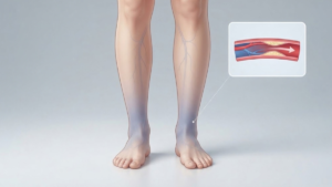
Chronic venous insufficiency is a long term condition in which leg veins fail to return blood to the heart efficiently. Blood pools in the lower leg, and pressure inside the vein stays high. Doctors call that raised pressure venous hypertension.
The failure can involve the surface veins, the deep veins, or the perforating veins that connect the two. Many patients have trouble in more than one system. Because pressure drives the damage, the condition rarely holds still. Swelling comes first for most people. Skin changes follow years later. In advanced cases, an open ulcer forms near the ankle. CVI often looks cosmetic at the start, yet it is a circulation problem from day one.
How Leg Veins Are Supposed to Work
One-Way Valves
Leg veins contain thin flaps of tissue spaced along their length. These valves open upward to let blood pass toward the heart. They then close behind it, so blood cannot slide back down. A single vein may hold dozens of them. Valves in the deep veins carry the heaviest load, because those vessels return roughly ninety percent of the blood leaving the leg. When a valve stays open or fails to seal, the column of blood above it presses straight back down.
The Calf Muscle Pump
Your calf muscles act as a second heart for the legs. Each step squeezes the deep veins and drives blood upward. The valves then lock that gain in place. Together, the two systems drop vein pressure sharply during walking. Standing still cancels the benefit. So does a stiff ankle, which explains why patients with arthritis or limited motion often swell more. Movement, in short, is part of the circulation itself.
What Goes Wrong in CVI: Venous Reflux Explained
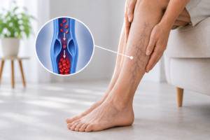
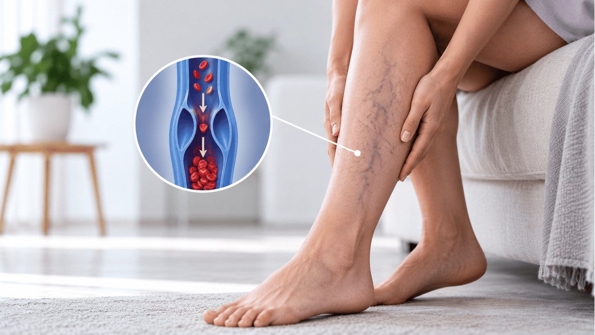
Venous reflux is the core problem in CVI. The term simply means blood flowing the wrong way inside a vein.
When a valve fails, blood falls backward every time you stand. The vein below it holds more blood than it should. That extra volume stretches the vein wall, which pulls the next valve apart. One leaking valve therefore creates another, and the damage spreads down the leg.
High pressure then forces fluid, proteins, and red blood cells out of the capillaries into surrounding tissue. Swelling follows. Iron released from those cells stains the skin brown. Inflammation sets in and slowly damages the tissue. Reflux explains nearly every symptom of CVI, from evening heaviness to open ulcers.
What Causes Chronic Venous Insufficiency?
Several distinct problems produce the same end result. Your treatment depends on which one applies to you.
Damaged or Weakened Vein Valves
Primary valve failure is the most common cause. The vein wall weakens first, often for genetic reasons. As the vessel widens, the valve leaflets no longer meet in the middle, so blood leaks past them. No clot or injury is involved. Doctors call this primary venous insufficiency, and it accounts for most cases seen in clinic. Age, hormone exposure, and years of standing all speed the process.
Prior Deep Vein Thrombosis (Post-Thrombotic Syndrome)
A clot in a deep vein damages every valve it touches. Even after the clot clears, scar tissue leaves the vein stiff and narrowed. Flow slows, and reflux begins. Doctors call this pattern post thrombotic syndrome, and it develops in a substantial share of patients within a few years of a deep vein thrombosis. Symptoms run more severe than in primary disease, and ulcers appear earlier. Anyone with a clot history should mention it at the first visit.
Vein Compression, Including Iliac Vein Compression
Sometimes the valves work fine and the outflow is blocked instead. The left iliac vein runs beneath the right iliac artery in the pelvis. In many people the artery presses on it and narrows the channel, a pattern known as May Thurner syndrome. Blood then backs up into the left leg. Suspect compression when only one leg swells, when swelling reaches the thigh, or when standard vein treatment fails to help. Imaging of the pelvis settles the question.
Genetics, Pregnancy, Obesity, and Prolonged Standing
Family history is the strongest single predictor of vein disease, and risk climbs sharply when both parents are affected. Pregnancy softens the vein wall through hormones and adds pressure from the growing uterus, so each additional pregnancy raises risk further. Excess abdominal weight keeps pelvic vein pressure high all day. Jobs that demand hours on your feet, such as nursing, teaching, and retail work, sustain that pressure for decades. These factors rarely act alone.
Who Gets CVI? Risk Factors and Prevalence
CVI is far more common than most people assume. Population studies in Western countries suggest that visible varicose veins affect roughly one in four adults. Advanced skin changes appear in a smaller group, and active venous ulcers affect close to one percent of adults. Prevalence rises with every decade of life.
Women report symptoms more often, partly because of pregnancy and hormonal effects. Men, however, are more likely to reach the ulcer stage before they seek care. Other risks include a previous leg fracture or surgery, smoking, and a sedentary routine. Height plays a small role too, since taller people carry a longer column of blood above the ankle.
The Signs and Symptoms of CVI
Symptoms build over years, so many patients adapt without noticing. A useful clue is timing. Vein symptoms grow worse through the day and settle overnight, while artery problems behave differently.
Common complaints include the following:
- Heaviness, aching, throbbing, or fatigue in the legs
- Ankle swelling that eases by morning
- Night cramps, restless legs, or deep sock marks
- Itching or burning over a bulging vein
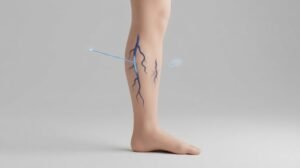
- Spider veins, or thick ropey varicose veins
- Brown discoloration and dry, flaking skin near the ankle
Heat and long periods of standing make everything worse. Walking and leg elevation bring relief. Later signs point to tissue damage rather than simple congestion. Firm skin that feels bound down, white scarred patches, and an open sore above the inner ankle all mark advanced disease and deserve prompt evaluation.
The 6 Stages of CVI (CEAP Classification)
Vascular specialists grade severity with the CEAP system. The clinical score runs from C0 through C6, and it guides both treatment planning and insurance approval.
C0 to C1: No Signs to Spider Veins
C0 means no visible signs at all. Patients may still feel aching or heaviness, and ultrasound may still show reflux. C1 covers spider veins and small reticular veins, the fine blue or red lines just under the skin. Both stages usually respond to compression and daily activity. Treatment here is often elective, although the symptoms themselves still deserve evaluation.
C2 to C3: Varicose Veins and Swelling
C2 describes true varicose veins, defined as bulging veins wider than three millimeters while standing. C3 adds visible swelling with no skin damage yet. Most patients finally seek help at this point. It is also the ideal moment to intervene, because closing the refluxing vein now prevents the skin changes that otherwise follow.
C4: Skin Changes and Discoloration
C4 marks the start of permanent damage. Brown staining, known as hemosiderin deposition, appears above the ankle. Venous eczema brings dry, itchy, flaking patches. In more advanced C4 disease the tissue hardens and the ankle narrows, a change called lipodermatosclerosis. White scarred areas may show up as well. These changes rarely reverse completely, so treatment should not wait.
C5 to C6: Healed and Active Venous Ulcers
C5 means an ulcer has healed while the skin stays fragile. C6 means an ulcer is open now. Venous ulcers sit most often just above the inner ankle. They look shallow and irregular, and they cause less pain than their appearance suggests. Healing takes months with compression alone, and recurrence is common unless the underlying reflux is corrected.
Is CVI the Same as Varicose Veins?
No, although the two overlap constantly. Varicose veins are a visible sign. CVI is the underlying failure of blood return that often produces them.
You can have one without the other. Some patients show prominent varicose veins with normal pressure and few symptoms. Others have severe deep vein reflux, heavy swelling, and no bulging veins on the surface at all. Body weight also hides surface veins, which delays diagnosis in many patients. Ultrasound settles the question, because it measures flow rather than appearance.
CVI vs. DVT: Two Different Vein Problems
Patients confuse these two often, yet they behave very differently. Deep vein thrombosis is a blood clot. It develops suddenly, usually in one leg, and it brings pain, warmth, redness, and firm swelling over hours or days. DVT is a medical emergency, since part of the clot can travel to the lungs.
CVI develops over years instead. It affects both legs in most patients, it improves with elevation, and it does not cause fever or sudden pain. The two conditions do connect, though. A past DVT is a leading cause of CVI. If one leg swells suddenly, seek care the same day.
How Is CVI Diagnosed?
Diagnosis begins with your history and a physical exam performed while you stand. Standing fills the veins and reveals reflux that hides when you lie flat.
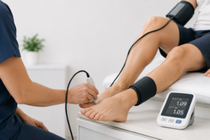
Venous duplex ultrasound is the standard test. A venous duplex scan combines an image of the vein with a measurement of flow. The technologist compresses your calf, releases it, and times how long blood runs backward. Reflux beyond half a second in a surface vein, or one second in a deep vein, confirms the diagnosis. The scan also maps which veins are involved and rules out a clot.
If pelvic vein compression seems likely, your physician may add CT venography, MR venography, or intravascular ultrasound for a closer look.
How Is CVI Treated?
Treatment lowers vein pressure, relieves symptoms, and protects the skin. Care follows a ladder that starts with the least invasive step.
Compression and Conservative Therapy
Graduated compression stockings come first for nearly everyone. They squeeze hardest at the ankle and support the calf pump. Most patients use 20 to 30 mmHg, while higher grades treat ulcers. Put them on before you rise in the morning. Pair them with daily walking, ankle exercises, leg elevation above heart level, and weight control. Conservative care manages symptoms well, though it cannot repair a failed valve.
Vein Ablation
Vein ablation closes the diseased vein from the inside. A catheter delivers heat from radiofrequency or laser energy, or the physician injects a medical adhesive that seals the vessel. The procedure takes under an hour, uses local anesthesia, and lets you walk out the same day. Blood immediately reroutes through healthy veins, so circulation improves rather than worsens. Published closure rates exceed ninety percent at long term follow up.
Sclerotherapy
Sclerotherapy treats smaller veins that a catheter cannot reach. The physician injects a liquid or foam solution that irritates the vein lining and causes it to seal shut. Foam works especially well for larger tributaries and for veins feeding an ulcer. Sessions run fifteen to thirty minutes, and several rounds are often needed. Bruising and temporary discoloration are the usual side effects, and both fade over a few weeks.
Stenting for Vein Compression
When a pelvic vein is narrowed, opening it corrects the outflow problem directly. During stent placement, the physician passes a catheter through a small puncture and positions a stent that holds the vein open. Patients with iliac vein compression often notice a fast drop in swelling and heaviness. Blood thinning or antiplatelet therapy follows for a set period, and ultrasound checks the stent over time.
Can CVI Be Cured or Reversed?
Partly. Doctors can close or bypass the faulty veins and bring pressure back toward normal. Symptoms such as aching and swelling often resolve completely. Ulcers heal and stay healed far more reliably once reflux is corrected.
The underlying tendency remains, however. Vein walls that weakened once can weaken again in other vessels. Skin changes from stage C4 rarely fade entirely, because the tissue has already scarred. So the honest answer is this. CVI is highly treatable and well controlled, yet it calls for lifelong attention.
What Happens If CVI Is Left Untreated
Untreated CVI does not stay the same. Pressure keeps rising, and the disease climbs the CEAP ladder. Swelling turns constant rather than evening only. Skin darkens and hardens. Venous eczema itches badly, and scratching breaks the surface, so minor knocks become wounds that will not close.
Other complications include bleeding from a thin surface vein, which can be sudden and heavy, and superficial thrombophlebitis, a painful clot inside a bulging vein. Repeated skin infection, or cellulitis, also becomes more likely. Roughly one in twenty patients with vein disease eventually develops an ulcer. Sleep, mobility, and work capacity all suffer along the way.
When to See a Vascular Specialist
Do not wait for an ulcer. Book an evaluation if leg swelling lasts more than a few days, if aching limits your routine, or if the skin near your ankle changes color or texture.
Come in sooner if you have a clot history, if only one leg swells, or if a varicose vein bleeds. Seek emergency care for sudden painful swelling in one leg. The team at Vascular Care Centers evaluates these symptoms in a single visit, and the ultrasound itself is quick and painless.