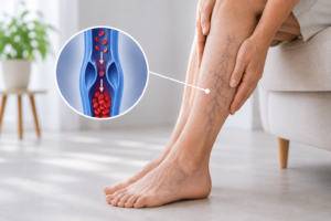
Leg swelling usually affects both sides. When only the left leg swells, the cause often sits higher up, inside the pelvis. Iliac vein compression syndrome is a common reason, and many patients carry it for years without a diagnosis.
Doctors also call it May-Thurner syndrome. An artery presses on a vein deep in the pelvis, and blood then struggles to leave the leg. Standard vein treatment does not fix the problem, because the blockage sits above the veins being treated. This article explains the anatomy, the warning signs, and the procedure that corrects it.
What Is Iliac Vein Compression Syndrome?
Iliac vein compression syndrome describes a narrowing of the iliac vein caused by pressure from a nearby artery. That vein carries blood from the leg and pelvis back toward the heart. When it narrows, blood backs up into the leg.
The compression itself is anatomical and present from birth in most people. Trouble starts only when the narrowing grows severe, or when the vein wall reacts to constant pressure. Over years, scar tissue forms inside the vein and creates fixed webs or spurs. Flow then drops further.
Not everyone with compression develops symptoms. Imaging studies show mild narrowing in a large share of healthy adults. Doctors therefore reserve the word syndrome for patients who have both the narrowing and matching symptoms.
May-Thurner Syndrome vs. Iliac Vein Compression: Same Condition?
For practical purposes, yes. The two terms describe the same problem, although they carry slightly different histories.
May and Thurner published the classic description in 1957 after examining cadavers. They found fibrous spurs inside the left common iliac vein at the point where the right iliac artery crossed over it. Their names stayed attached to that specific pattern.
Iliac vein compression syndrome is the broader label. It also covers narrowing at other points, such as the right iliac vein, or a vein squeezed by a tumor, a full bladder, or a pregnant uterus. Older papers use the term Cockett syndrome as well. Vascular specialists treat these names as interchangeable in daily practice.
How the Right Artery Compresses the Left Vein
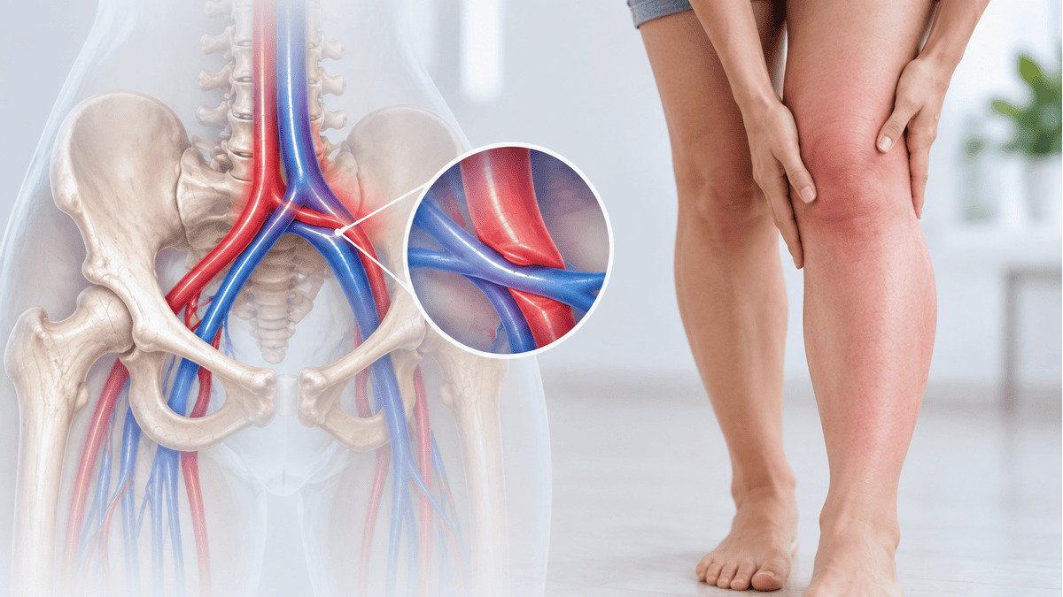
The anatomy explains everything. Blood leaving each leg travels up through the common iliac vein. The left and right veins then join to form the inferior vena cava, which sits slightly to the right of the spine.
Because of that position, the left iliac vein must cross the midline. It passes underneath the right common iliac artery along the way, and the spine lies directly behind it. The vein therefore sits pinned between a pulsing artery in front and bone at the back.
Every heartbeat sends a pulse through that artery. Over decades, the repeated pressure irritates the inner lining of the vein. Cells respond by laying down fibrous tissue. The result is a fixed narrowing that no longer opens, even when the patient lies down.
Why Symptoms Almost Always Affect the Left Leg
The crossing point sits on the left, so the left leg drains against the highest resistance. Roughly nine out of ten symptomatic cases involve the left side.
Right sided compression does occur, though far less often. It usually follows an unusual course of the arteries or pressure from a mass inside the pelvis.
This left sided pattern gives doctors a useful clue. Swelling in both legs points toward the heart, kidneys, liver, or general vein disease. Swelling limited to the left leg, especially in a younger patient, should always raise the question of pelvic vein compression.
Symptoms of Iliac Vein Compression
Symptoms range from mild to disabling. Many patients notice a slow change across years, while others present suddenly with a blood clot.
One-Sided Leg Swelling
Swelling in one leg is the hallmark sign. It usually begins in the ankle and calf, then spreads to the thigh as the condition progresses. The leg feels tight by evening and improves overnight at first. Later, the swelling stays all day.
Patients often notice that one shoe or one pant leg fits differently. Measuring both calves at the same level makes the difference obvious. A gap of more than two centimeters deserves investigation.
Pelvic Pain and Heaviness
Raised pressure inside the pelvic veins causes a dull ache low in the abdomen, the groin, or the buttock. Women may report pain that worsens before a period, during intercourse, or after long periods of standing. Some develop visible varicose veins around the vulva or the upper inner thigh.
A related symptom is venous claudication. The leg feels bursting or tight after several minutes of walking, and relief comes with rest and elevation.
Varicose Veins That Keep Coming Back
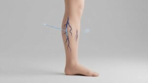
Recurrence after sound vein treatment is a red flag. If varicose veins return quickly on the left side after ablation or surgery, the outflow above them may still be blocked.
The same applies when veins appear in unusual places, such as across the lower abdomen or the pubic area. Those vessels act as detours around the narrowing. Treating them without opening the iliac vein rarely holds.
Sudden DVT in a Young, Healthy Person
Iliac vein compression is a leading cause of deep vein thrombosis in young adults, especially women between twenty and fifty. The clot typically fills the left leg from the groin downward.
Suspect compression when a clot appears without a clear trigger, when it affects the left leg, or when it returns despite blood thinners. Sudden painful swelling with warmth and redness needs same day medical care.
Who Is Most Commonly Affected?
Women account for the majority of diagnosed cases. Most present between the ages of twenty and fifty.
Several factors raise the risk. Pregnancy adds pressure from the uterus and softens the vein wall through hormones. Birth control containing estrogen increases clotting risk. Long periods of immobility, recent surgery, dehydration, and inherited clotting disorders all add to the load.
Anatomy still comes first, however. Autopsy and imaging studies show a meaningful degree of compression in a large minority of the general population. Most of those people never develop symptoms, so screening healthy adults is not recommended.
The Link Between Iliac Vein Compression and Chronic Venous Insufficiency
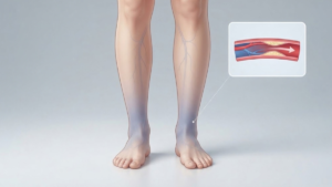
Chronic venous insufficiency usually begins with leaking valves in the leg veins. Iliac vein compression produces the same end result through a different route. Instead of blood falling backward, blood cannot get out. Either way, pressure inside the leg veins stays high. That pressure drives swelling, skin staining, and eventually ulcers.
The two problems often coexist. High pressure from the pelvis stretches the veins below it, which pulls the valves apart and creates reflux. Treating only the leg veins leaves the source untouched. For that reason, specialists look at the pelvis whenever symptoms outrun the ultrasound findings.
Why This Condition Is Frequently Missed
Standard leg ultrasound stops at the groin in many labs. The pelvic veins sit deep behind the bowel, so they are harder to see. A normal leg scan therefore does not rule out compression.
Symptoms also mimic other conditions. Doctors may attribute the swelling to weight, lymphedema, or ordinary varicose veins.
Access plays a part too. Many vein clinics treat surface veins but do not perform pelvic venography or stenting, so the diagnosis never enters the conversation. Patients often move through several providers before someone images above the groin.
How Iliac Vein Compression Is Diagnosed
Diagnosis combines symptoms with imaging. No single test answers every question, so physicians usually order these studies in sequence.
Duplex Ultrasound
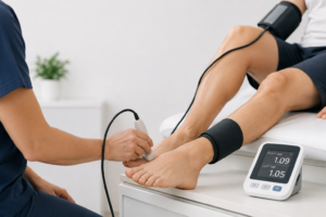
A venous duplex scan is the first step. It checks the leg veins for clots and reflux, then examines the pelvic veins where the view allows. Certain findings suggest compression even when the vein itself stays hidden. Blood flow that runs steady instead of changing with breathing points to a blockage above. A large difference in flow between the two sides carries the same meaning.
CT or MR Venography
Cross sectional imaging shows the pelvis clearly. CT venography uses contrast dye and gives a fast, detailed picture of the artery crossing the vein. MR venography avoids radiation, which suits younger patients and repeat studies.
Both tests measure how far the vein narrows. Remember that the patient lies flat during the scan, so compression can look milder than it does during standing or walking.
Intravascular Ultrasound (IVUS)
IVUS is the reference standard. A tiny ultrasound probe travels inside the vein on a catheter and images the wall from within. The probe measures the true cross sectional area and reveals scar tissue that outside imaging misses. Most specialists treat a reduction in area beyond fifty percent as significant. IVUS also guides stent sizing and confirms a good result before the procedure ends.
Treatment: Iliac Vein Stenting
Opening the vein is the definitive treatment. The procedure is minimally invasive and works through a small puncture, usually behind the knee or in the groin.
The physician passes a catheter to the narrowed segment, widens it with a balloon, then places a self expanding stent designed for veins. Venous stents are wider and more flexible than arterial stents, since they must resist steady outside pressure. IVUS then confirms full expansion.
Stent placement takes about an hour. Most patients go home the same day and start a blood thinner or an antiplatelet drug afterward.
When a fresh clot sits in the vein, treatment begins with thrombectomy or thrombolysis to clear it. The stent goes in once flow returns.
What Recovery Looks Like After Venous Stenting
Recovery is quick for most patients. Walking begins the same day, and desk work usually resumes within two or three days. Avoid heavy lifting for about a week.
Many people notice less swelling within days. Aching and heaviness improve over the following weeks, though skin changes take longer to settle.
Follow up includes ultrasound at set intervals to confirm the stent stays open. Long term results are good, and most published series report that eighty to ninety percent of stents remain open several years later. Compression stockings and regular walking protect that result.
What Happens If It Goes Untreated
Untreated compression rarely stays stable. Pressure keeps building, and the leg pays the price.
Deep vein thrombosis becomes more likely, and a clot in the pelvis carries a real risk of traveling to the lungs. Patients who recover from a large clot often develop post thrombotic syndrome, with lasting swelling, pain, and skin damage.
Chronic swelling then moves through the familiar stages of vein disease. Skin darkens near the ankle, hardens, and eventually breaks down into an ulcer. Walking distance shrinks along the way. Early treatment prevents most of this.
If one leg swells more than the other, or if varicose veins keep returning after treatment, ask for imaging above the groin. The team at Vascular Care Centers evaluates pelvic vein compression directly, so the diagnosis does not depend on a leg scan alone.