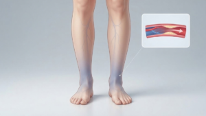
Two patients can walk into a clinic with identical looking legs and leave with very different
treatment plans. The difference comes from testing. Chronic venous insufficiency is a problem
of pressure and flow, and neither of those shows up on the surface.
A proper CVI workup follows a set order. Your physician takes a history, examines you while
you stand, then measures blood flow with ultrasound. Extra tests follow only when the picture
stays unclear. This article walks through each step, explains what the numbers on your report
mean, and covers what insurers usually ask for before they approve treatment.
Why CVI Requires Testing, Not Just a Visual Exam
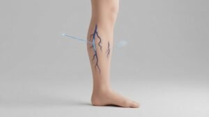
Visible veins tell you very little about severity. Some patients have thick, ropey varicose veins
with mild reflux underneath. Others have severe reflux, daily swelling, and almost nothing
showing on the skin.
The deep veins are the bigger blind spot. They carry roughly ninety percent of the blood leaving
your leg, and they sit far below the surface. No exam can assess them by sight or touch.
Testing also answers the questions that shape treatment. Which vein is leaking? How far does
the leak extend? Is anything blocking the outflow higher up? Is a clot present? A treatment plan
built without those answers usually fails, and insurers will not approve procedures without
documented findings.
Step 1: Medical History and Risk Assessment
Your first visit starts with questions, and the answers often point straight to the cause. Expect
your physician to ask how long symptoms have lasted, when they peak during the day, and
what makes them better.
Certain details carry extra weight. A previous deep vein clot suggests damaged valves and
scarring rather than simple valve weakness. Multiple pregnancies, a strong family history, and
years spent standing at work all raise the likelihood of primary vein disease.
Your physician will also review medications, since hormones raise clotting risk and several
common drugs cause swelling on their own. Questions about heart, kidney, liver, and thyroid
conditions help separate vein swelling from systemic swelling.
Many clinics score symptoms formally using the Venous Clinical Severity Score. That number
gives you and your physician a baseline to compare against after treatment.
Step 2: Physical Examination
The exam confirms what the history suggests and directs the ultrasound that follows.
Standing Exam and Vein Mapping
You will be asked to stand for several minutes before the exam begins. Standing fills the veins
under gravity and reveals bulges that vanish when you lie flat.
Your physician inspects the leg from the groin down to the foot, then feels along the course of
the major veins for firm cords or soft bulges. Veins in the inner thigh and calf usually follow the
great saphenous vein, while veins at the back of the calf follow the small saphenous vein. Some
clinicians mark the visible pattern on the skin before scanning, which makes the ultrasound
faster and more targeted.
Assessing Skin Changes
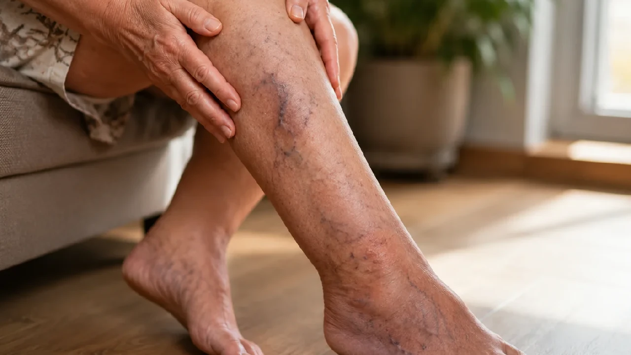
The skin above the ankle tells the story of how long pressure has been high. Your physician
looks for brown staining, dry and flaking patches, white scarred areas, and firm tissue that feels
bound down.
Swelling gets checked by pressing a thumb into the shin and watching how the dent refills. Both
calves are measured at the same level, since a difference of more than two centimeters points
toward a blockage on one side. Any ulcer is measured and photographed for the record.
CEAP Staging
CEAP is the standard classification for venous disease, and it has four parts. C stands for
clinical stage, running from C0 with no visible signs through C6 with an open ulcer. E covers the
cause, which may be primary valve failure, secondary damage after a clot, or a condition
present from birth.
A describes the anatomy involved, meaning the surface veins, the deep veins, the perforating
veins, or a combination. P describes the mechanism, which is reflux, obstruction, or both
together. Your final classification looks something like C4a, secondary, superficial and deep,
reflux and obstruction.
Step 3: Venous Duplex Ultrasound
Duplex ultrasound is the central test for CVI. It combines a live image of the vein with a
measurement of blood flow, so it shows both structure and function in one sitting. The scan
takes roughly forty five minutes per leg, uses no needles or dye, and causes no pain.
Why the Test Is Performed Standing
Gravity is the whole point. Lying down empties the leg veins and hides the very problem the test
is meant to find, so reflux measured on a table can look normal when it is not.
Most labs scan with the patient standing on a low platform, holding a support bar, with weight
shifted onto the opposite leg. Patients who cannot stand safely are tilted steeply on the table
instead, which reproduces some of the same effect. If your previous scan was done lying flat,
the results deserve a second look.
What Venous Reflux Time Means
Reflux time measures how long blood flows the wrong way inside a vein. The technologist
squeezes your calf to push blood upward, then releases quickly. In a healthy vein, the valve
snaps shut and backward flow stops almost at once.
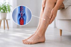
When a valve leaks, blood keeps falling back down, and the ultrasound tracks that reversed flow
as a waveform. The length of that backward flow, measured in seconds, is the number your
report will show for each vein segment.
The 500-Millisecond Threshold
Half a second is the accepted cutoff for the surface veins. Backward flow lasting longer than 500
milliseconds in a saphenous vein counts as abnormal, and shorter reversals fall within normal
limits.
The deep veins get a more forgiving threshold of one full second, because they normally carry
brief reversals during breathing and movement. Perforating veins, which connect the two
systems, are usually judged abnormal beyond about 350 milliseconds when they also measure
wide enough to matter.
Which Veins Get Mapped
A complete study covers the deep system, the surface system, and the connections between
them. Deep veins include the common femoral, femoral, and popliteal veins. Surface veins
include the great saphenous, the small saphenous, and the anterior accessory saphenous vein.
For each segment, the technologist records whether the vein compresses fully, which rules out
a clot, plus its diameter in millimeters and its reflux time. Diameter matters because insurers
often require a minimum size before approving treatment, and because a wider vein tends to
leak more.
Additional Tests When Duplex Isn’t Conclusive
Sometimes symptoms outrun the findings. When swelling is severe and the leg ultrasound looks
reasonable, the problem often sits higher up.
CT or MR Venography
These scans image the pelvis and abdomen, where a standard leg ultrasound cannot reach
well. They show the iliac veins, the inferior vena cava, and any mass pressing on them. CT
venography uses contrast dye and produces fast, detailed images. MR venography avoids
radiation, which suits younger patients and repeat studies. Remember that both are performed
lying flat, so compression can appear milder than it feels.
Intravascular Ultrasound
Intravascular ultrasound, shortened to IVUS, places a small probe inside the vein on a catheter
and images the wall from within. It is the reference standard for pelvic vein narrowing, since it
measures the true cross sectional area and reveals scar tissue that outside imaging misses.
Most specialists treat a reduction in area beyond fifty percent as significant.
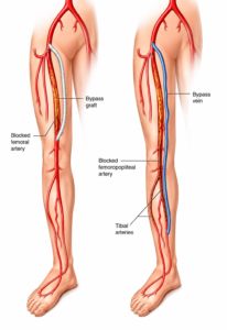
Ankle-Brachial Index to Rule Out Artery Disease
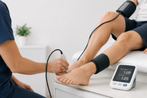
This quick test compares blood pressure at the ankle with blood pressure in the arm. A ratio
below 0.9 indicates reduced arterial flow. The result changes your treatment directly, because
firm compression can be unsafe when arterial supply is already poor. Very stiff, calcified arteries
can produce a falsely high reading, so a toe pressure measurement is sometimes added.
Ruling Out Other Causes of Leg Swelling
Vein disease is common, yet it is not the only reason legs swell. A careful workup rules out the
alternatives before settling on a diagnosis.
Heart, kidney, and liver disease produce swelling on both sides at once, usually with other clues
such as breathlessness or rapid weight gain. Low protein levels and an underactive thyroid do
the same. Several medications, including certain blood pressure drugs and steroids, cause
swelling as a side effect.
Lymphedema involves the foot and toes, which vein swelling tends to spare, and the tissue feels
firm rather than soft. Lipedema causes symmetric fatty enlargement that stops at the ankle.
Basic blood work sorts out most of these, and your physician orders it selectively rather than
routinely.
How to Read Your CVI Diagnosis Report
Ultrasound reports look technical, though the format is consistent. Each vein segment gets its
own line with three pieces of information.
Compressibility comes first. A vein described as fully compressible contains no clot. Diameter
comes next, listed in millimeters and usually measured while standing. Reflux time comes last,
given in seconds or milliseconds.
A typical line might read that the great saphenous vein is patent and compressible, measures
7.2 millimeters at the groin, and shows reflux of 1.8 seconds from the junction to the mid calf.
Translated, that vein carries no clot, is enlarged, and leaks well beyond the half second
threshold along most of its length.
Words such as competent and incompetent simply mean the valves work or they do not. Ask
your physician to walk through the report with you, since the segments listed determine which
procedures make sense.
What Your Diagnosis Means for Treatment
Findings map onto treatment fairly directly. Reflux limited to a surface vein responds well to
closing that vein, since blood reroutes through healthy vessels nearby.
Reflux confined to the deep veins is different. Those vessels cannot be closed, so care centers
on compression, exercise, elevation, and weight control, with treatment of any surface reflux
that adds to the load.
Obstruction calls for a different approach again. When imaging shows a narrowed pelvic vein,
opening it with a stent addresses the cause, and treating the leg veins alone tends to
disappoint. Your CEAP stage sets the urgency. Early stages allow a measured pace, while skin
changes and ulcers call for prompt treatment to prevent permanent damage.
How to Prepare for a Vein Evaluation
A little preparation makes the visit faster and the results more reliable. Bring any earlier
ultrasound reports, operative notes from previous vein procedures, and a current list of your
medications.
Wear loose clothing or shorts, since both legs need to be exposed from the groin down. Skip
lotion on the day of the scan, because it interferes with the ultrasound probe. Plan to stand for
part of the appointment, and set aside about an hour. No fasting is required, and drinking water
beforehand actually helps.
Write down your questions in advance and note when your symptoms are worst. The team at
Vascular Care Centers completes the history, exam, and duplex ultrasound in a single visit, so
most patients leave with a diagnosis and a plan the same day.