Procedures We Perform
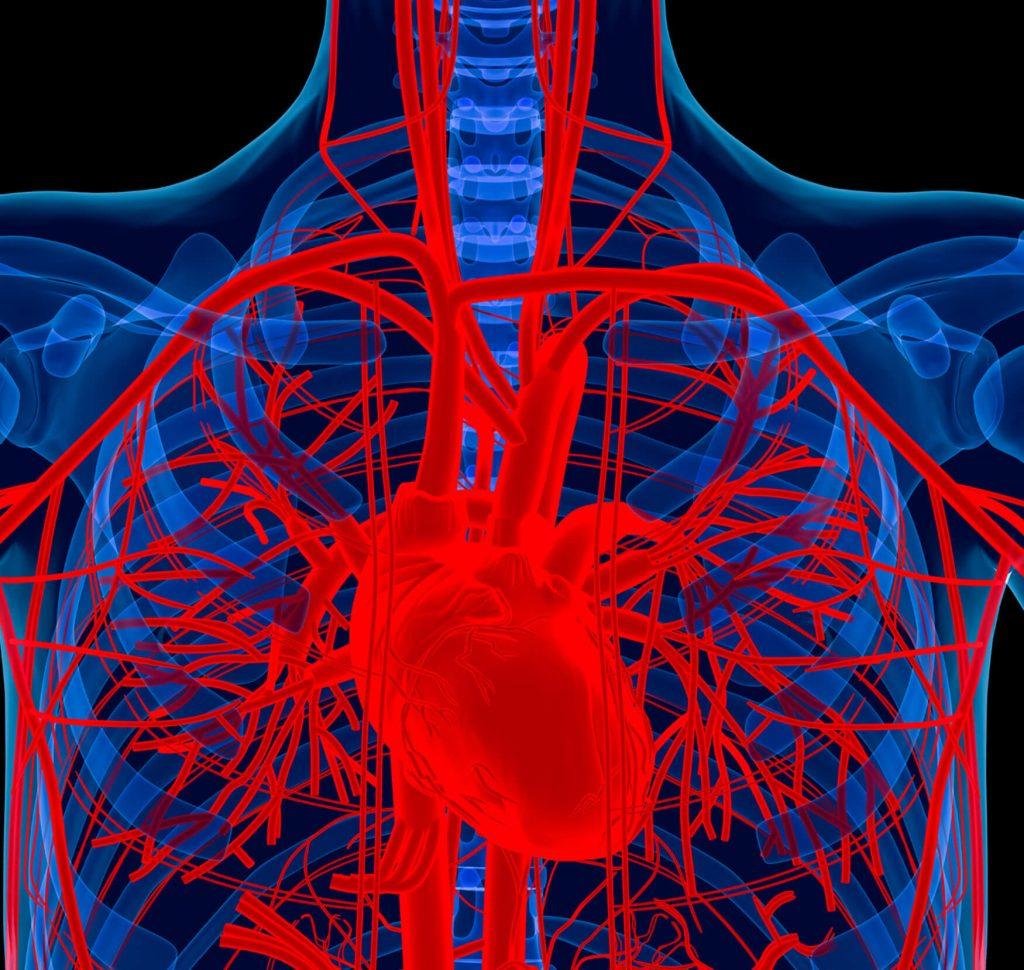
A Vascular Care Centers specializes in diagnosing and treating blood vessel conditions using advanced procedures like angioplasty, stent placement, arterial duplex ultrasound, and venous ablation.Our expert team provides minimally invasive treatments to restore healthy blood flow and improve overall vascular health.
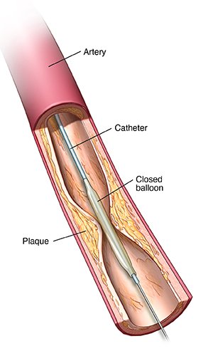
Angioplasty
Angioplasty is a minimally invasive procedure used in advanced Vascular Treatments to open narrowed or blocked arteries. It plays an important role in effective Artery Disease Treatment, helping restore proper blood flow, relieve pain, and improve overall circulation.
Procedure:
- Preparation: Your doctor reviews your medical history and medications. Local anesthesia is given, and ECG monitors your heart.
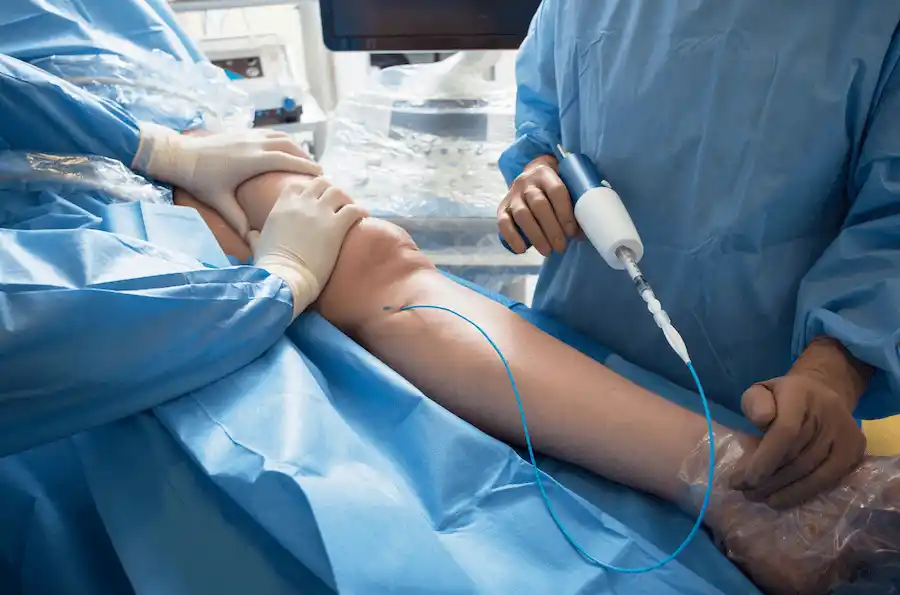
- Catheter Insertion: A thin catheter with a balloon is inserted into a blood vessel, usually through the groin, arm, or wrist.
- Locate Blockage: Contrast dye is injected, and X-ray imaging guides the catheter to the blocked artery.
- Open the Artery: The balloon is inflated to widen the narrowed artery, improving blood flow.
- Finish & Monitor: The balloon is deflated, catheter removed, and the site is closed. You rest while your condition is monitored for any complications.
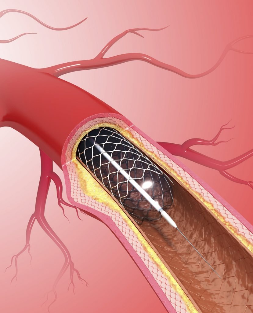
Stent Placement
Stents are small metallic mesh tubes designed to keep narrowed or blocked passageways in the body open. Different types of stents are used for various medical purposes, and they have transformed the treatment of conditions like coronary heart disease and other blockages by reducing the need for invasive surgery. Stents can be placed in coronary arteries to improve blood flow to the heart, cerebral arteries for brain circulation, or renal arteries to support kidney function. They come in various sizes to match the diameter of the affected blood vessel.
Procedure:
- Preparation: You lie on the table, and a health line (IV) is inserted. Local anesthesia is given, and pain management keeps you relaxed.
- Catheter Insertion: A thin catheter is inserted into a blood vessel, usually in the arm or groin.
- Locate Blockage: Using X-ray imaging, the catheter is guided to the blocked vessel, and contrast dye is injected to identify the blockage.
- Open the Artery: The balloon at the tip of the catheter is inflated to widen the vessel, and a stent is placed to keep it open.
- Finish & Monitor:The catheter is removed, and the insertion site is closed with sutures or manual pressure.
Arterial/Venous Duplex
Duplex Vascular Ultrasound is a safe, noninvasive diagnostic test used to evaluate blood flow in the arteries and veins. It uses sound waves to create real-time images and detect blockages, clots, or circulation problems. The procedure is painless, requires no anesthesia, and is performed on an outpatient basis. Patients can resume normal activities immediately after the test.
Procedure:
- Preparation: You lie comfortably on the examination table, and your blood pressure may be checked.
- Apply Gel:A clear gel is applied to the area being examined to help transmit sound waves.
- Use Transducer:A handheld device (transducer) is placed on your skin and moved over the arteries and veins.
- Image & Sound Capture: The transducer emits sound waves, which reflect back to create real-time images of blood flow; soft whooshing sounds may be heard.
- Evaluation:The images are reviewed by a vascular specialist and may be recorded for further analysis.
Venous Ablation
Venous ablation is a minimally invasive procedure used to treat varicose veins. This technique employs radiofrequency or laser energy to close and seal the affected veins. It helps relieve common symptoms of varicose veins, including pain, swelling, leg heaviness, and skin irritation.
Procedure:
- Preparation: Lie on the procedure table wearing a gown and protective glasses; the doctor uses ultrasound to locate the abnormal vein.
- Apply Gel: A catheter is inserted into the vein under sterile conditions.
- Anesthesia: Local anesthesia is applied at the insertion site and along the vein to ensure comfort.
- Vein Closure: A laser fiber or radiofrequency electrode is advanced through the catheter to close the vein, monitored continuously with ultrasound.
- Finish Procedure:The catheter is removed, the insertion site is covered with a bandage, and the patient can go home the same day.
Fistulogram
A fistulogram is a diagnostic imaging procedure used to evaluate the size, shape, and pathway of a fistula. A fistula is an abnormal connection between two body parts or passageways. These commonly occur between hollow organs such as the vagina, bladder, colon, or rectum.
Procedure:
- Preparation: You lie comfortably on the radiology table, and an IV line may be placed if medications or fluids are needed.
- Clean & Numb Area: The area around the fistula is cleaned with antiseptic, and local anesthesia may be given.
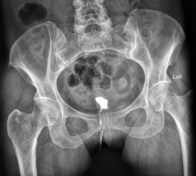
- Catheter Insertion: A thin catheter is gently inserted into the fistula under X-ray guidance.
- Imaging: Contrast dye is injected to outline the fistula, and images are captured while you may be asked to hold your breath briefly.
- Finish Procedure: The catheter is removed, and the insertion site is covered with a sterile dressing.
Atherectomy
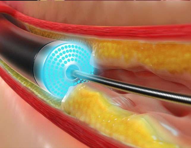
Atherectomy is a medical procedure designed to remove plaque buildup from blood vessels. Plaques are fatty deposits that can narrow arteries, increasing the risk of heart attack, stroke, and other serious complications. During atherectomy, a catheter equipped with a blade or laser is guided to the affected vessel to remove the plaque and restore healthy blood flow.
Procedure:
- Preparation: Local anesthesia is applied at the catheter insertion site (usually groin, arm, or wrist), and proper pain management keeps you comfortable while awake.
- Clean & Numb Area: A catheter is inserted and guided to the blocked artery using X-ray imaging.
- Catheter Insertion: Contrast dye is injected to create angiograms and identify the plaque; you may feel mild metallic taste or headache.
- Imaging: A blade or laser at the catheter tip removes the plaque, and the material is collected for evaluation.
- Finish Procedure: Blood flow is restored, the catheter is withdrawn, and the insertion site is closed with sutures or manual pressure.
Thrombectomy/ Thrombolysis
Thrombectomy/Thrombolysis is a medical procedure designed to remove or dissolve abnormal blood clots that block normal circulation and can damage vital organs or tissues. The procedure uses X-ray imaging to guide a catheter precisely to the site of the clot. It is commonly performed to reduce the risk of embolism, heart attack, and stroke, restoring proper blood flow and protecting organ function.
Procedure:
- Preparation: You lie comfortably on the procedure table while electrodes monitor your heart rate, blood pressure, oxygen levels.
- Catheter Insertion: A catheter is inserted into the artery under local anesthesia and sterile conditions.
- Locate Clot: The catheter is guided to the clot under X-ray guidance, and contrast dye is injected to pinpoint its exact location; you may notice a metallic taste or mild headache.
- Clot Dissolution: Clot-dissolving medication is delivered through the catheter, and in some cases, the clot is mechanically broken down.
- Finish Procedure: Once the clot is removed or dissolved, the catheter is withdrawn, and pressure is applied to the insertion site to prevent bleeding.

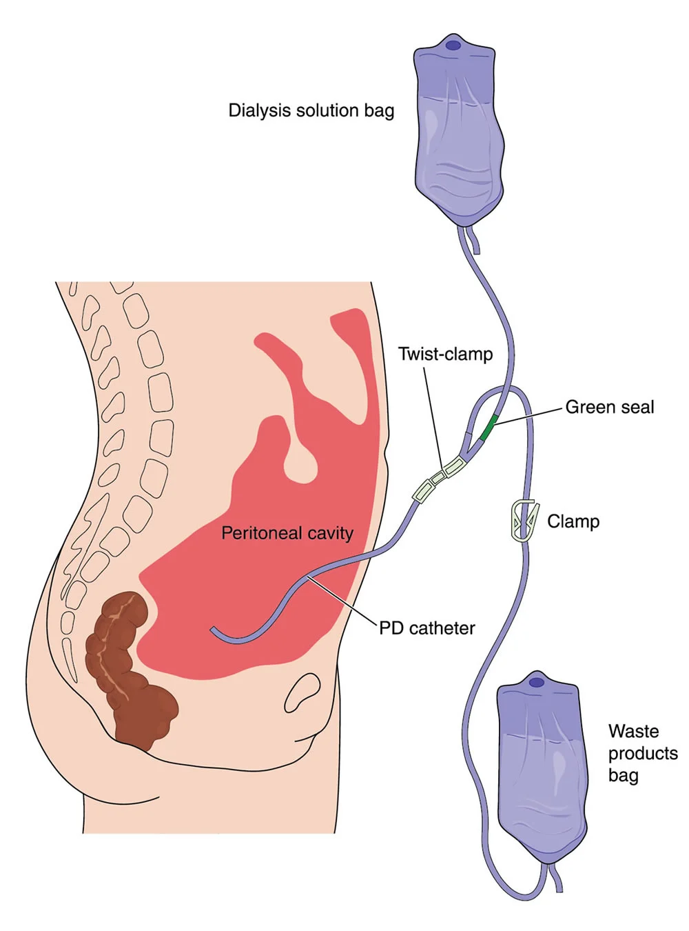
Dialysis Catheter Placement
A dialysis catheter is inserted to allow blood to flow from the body to a dialysis machine, where it is filtered and cleaned of waste products. Dialysis is necessary when the kidneys are no longer able to effectively remove toxins and excess fluid from the blood. A tunneled dialysis catheter provides reliable and immediate access to the bloodstream for ongoing treatment. When properly placed, it eliminates the need for repeated needle insertions during each dialysis session.
Procedure:
- Preparation: You lie comfortably on the procedure table, and local anesthesia is applied to numb the neck and chest areas.
- Access Vein: Small incisions are made, and a needle with a guidewire is used to access the jugular vein and advance into the vena cava.
- Create Tunnel: A tunnel is created beneath the skin between the neck and chest incisions to guide the catheter.
- Catheter Placement: The flexible dialysis catheter is inserted through the chest incision, guided by the guidewire, and positioned correctly in the vein; X-ray confirms placement and blood flow.
- Finish Procedure: The guidewire is removed, the catheter is secured with stitches, and both incision sites are closed and dressed to reduce infection risk.